THE CASE FILES

Complex Endodontic Case Studies

Real radiographs, real patients, real outcomes — the cases that show why dentists send us their hardest work. Including the ones where the right answer was no treatment at all.

15 DOCUMENTED CASES 5,000+ TEETH SAVED 100% REAL — NO STOCK PHOTOS
THE GALLERY

Every case, a painting.

Each frame holds a before and its after. Hover to see the transformation.
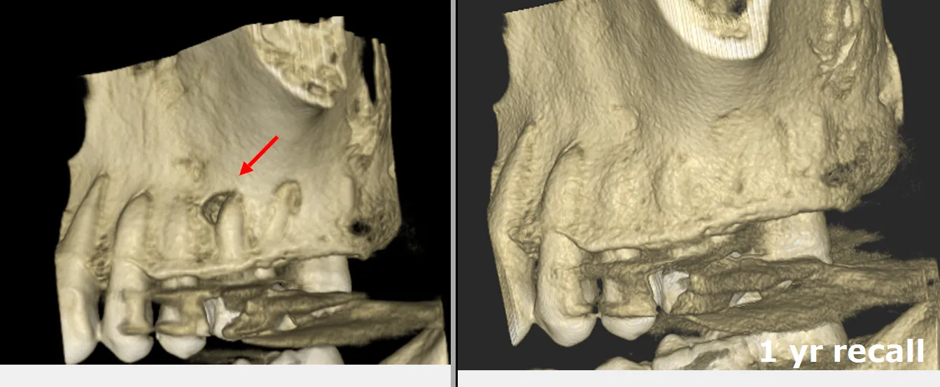
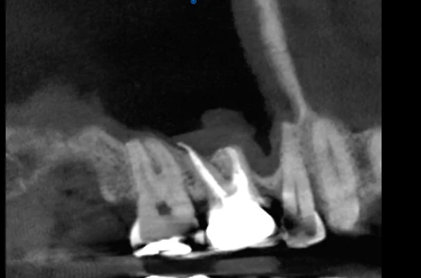
Recall CBCT showing complete healing and bone regeneration
Pre-operative CBCT showing missed MB2 canal and periapical bone loss
BEFORE AFTER
◂▸
CASE 001 — MAXILLARY FIRST MOLAR 2 HOURS The canal that wasn't there Two years of unexplained pain after a "finished" root canal — until 3D imaging revealed a fourth canal, sealed shut by calcification and invisible on every X-ray. COMPLEX ANATOMY ✦✦✦ EXTREME
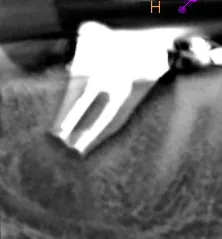
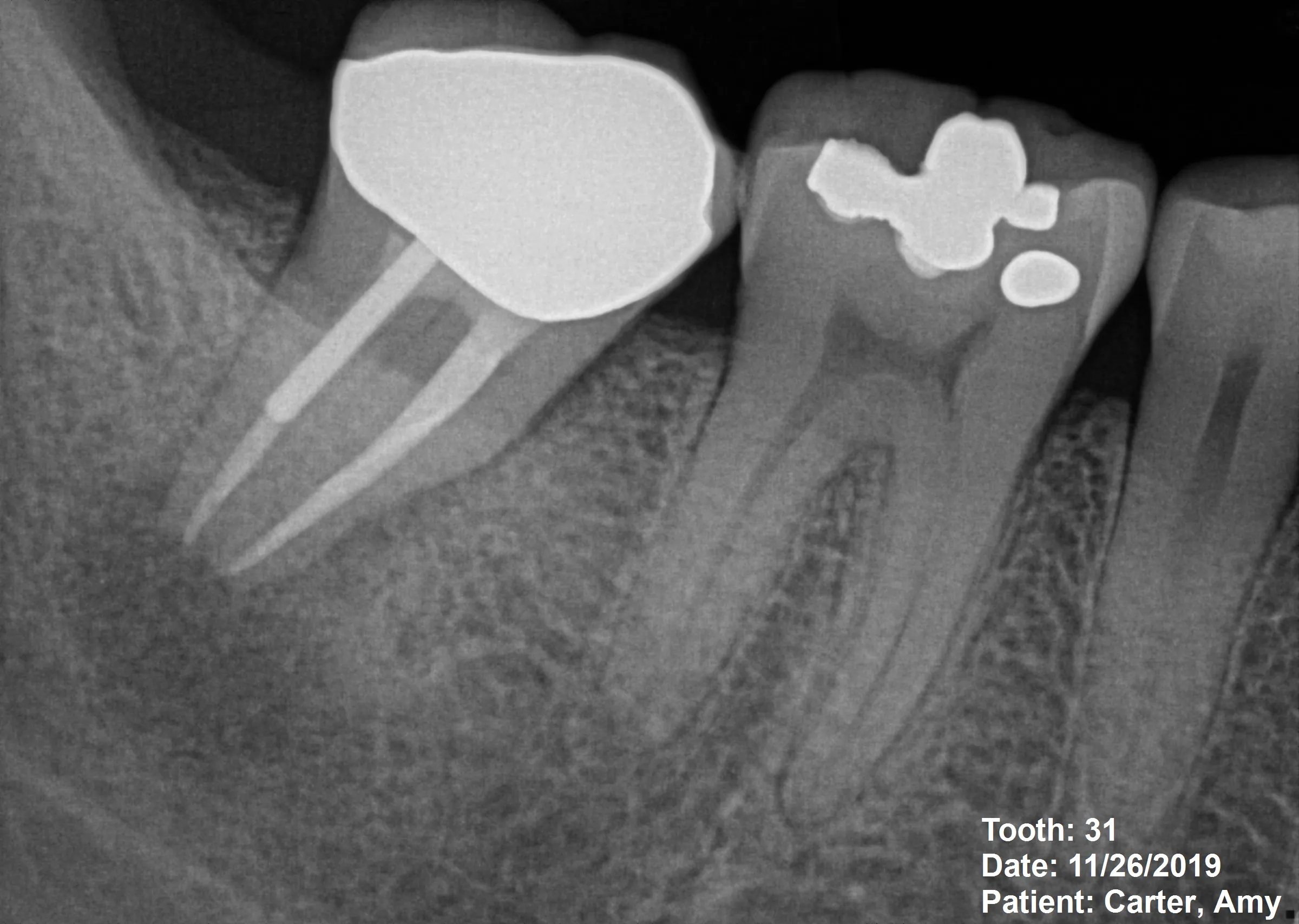
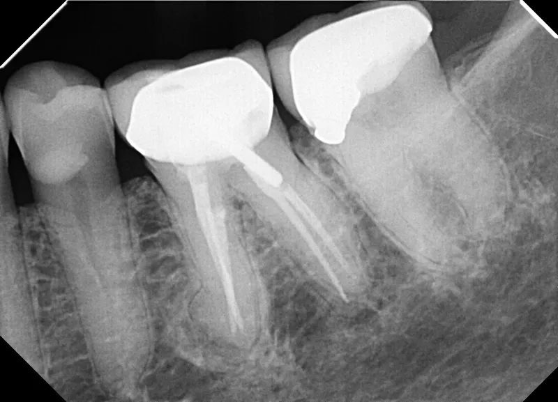
Final radiograph — complete three-dimensional obturation of the C-shaped anatomy
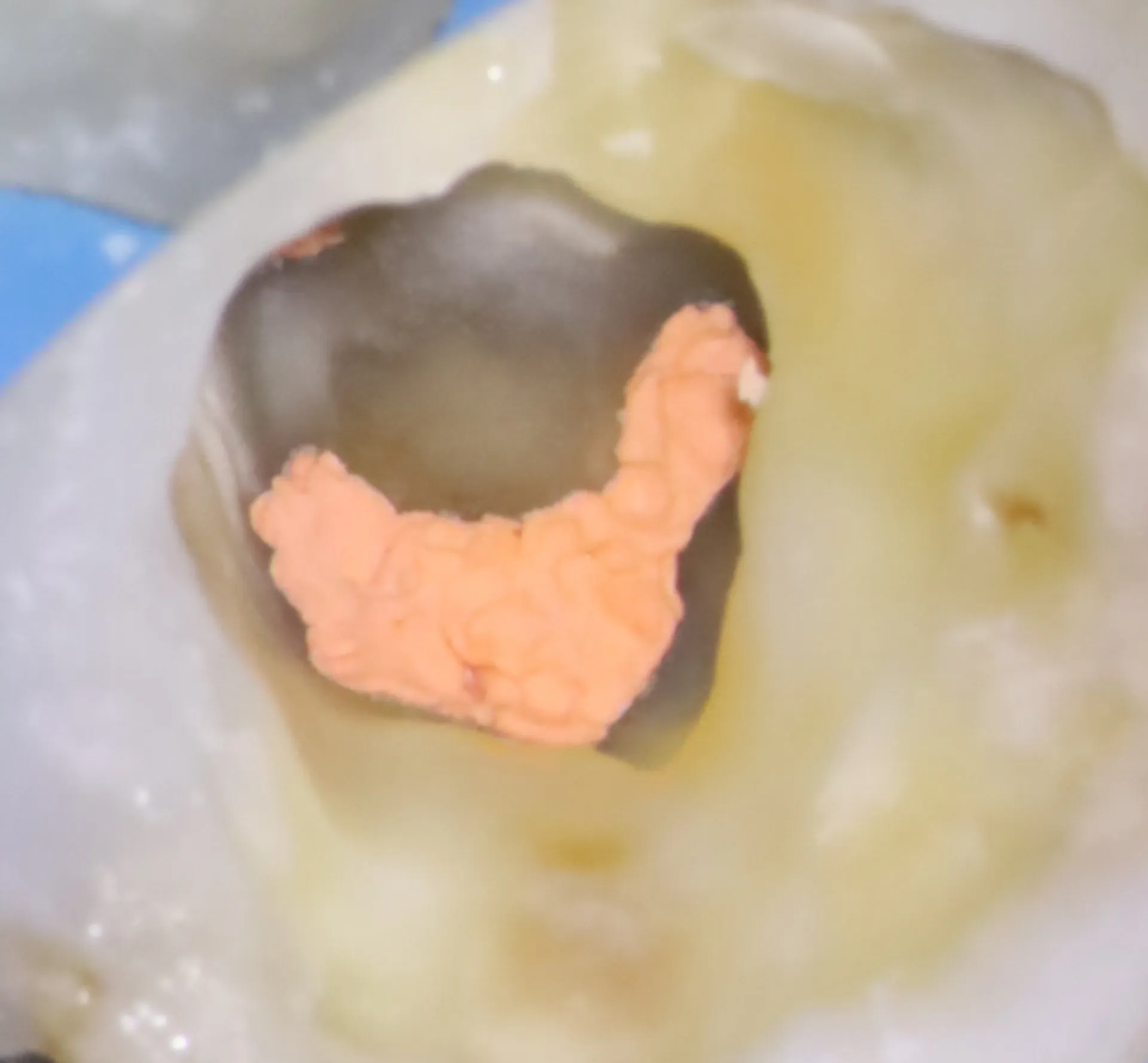
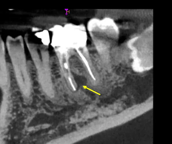
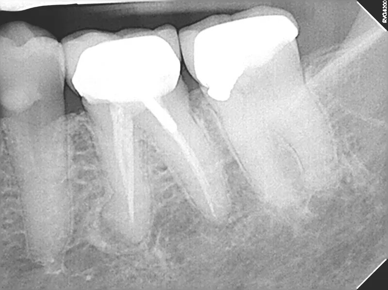
Pre-operative radiograph of mandibular second molar with severe decay
BEFORE AFTER
◂▸
CASE 002 — MANDIBULAR SECOND MOLAR 90 MINUTES The C-shaped labyrinth Severe decay in a lower molar hid a rarity: no separate canals at all, but one continuous C-shaped ribbon of pulp — anatomy that defeats every standard technique. COMPLEX ANATOMY ✦✦ CHALLENGING
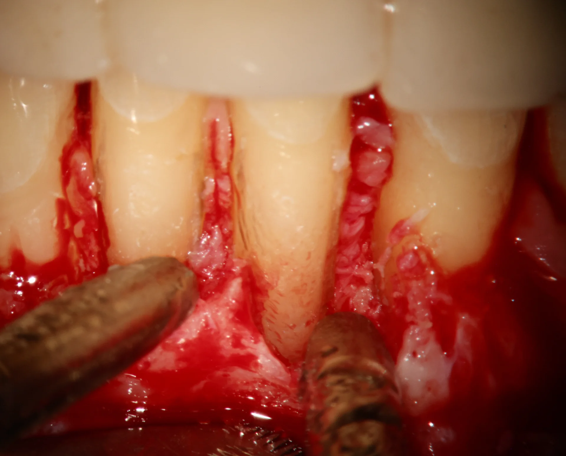
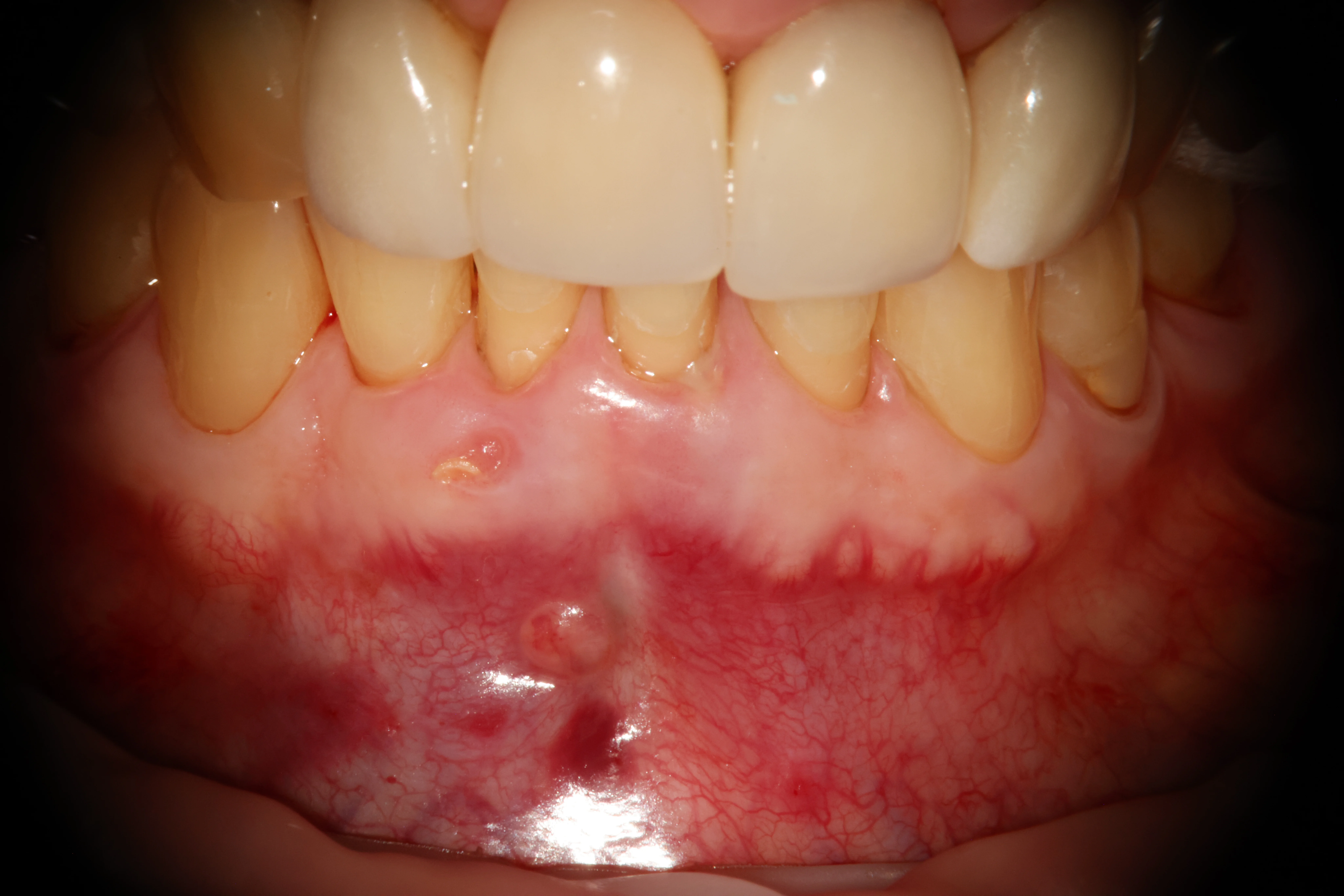
Post-surgical photo — sutured flap over the repaired cemental tear site
Pre-operative photo — buccal sinus tract near lower anterior teeth
BEFORE AFTER
◂▸
CASE 003 — TEETH #24 & #25 1 HOUR The tear beneath the surface A well-done root canal that wouldn't heal, a sinus tract that wouldn't close — because the real problem was a torn strip of cementum on the outside of the root, invisible to every X-ray. SURGERY ✦✦✦ EXTREME
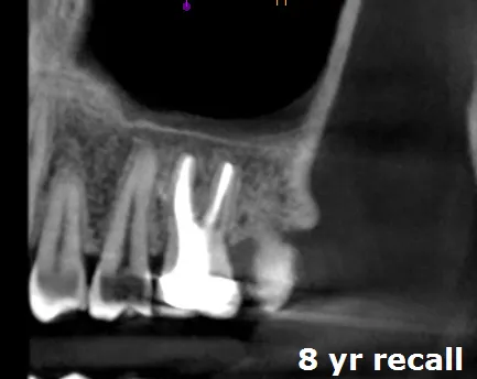
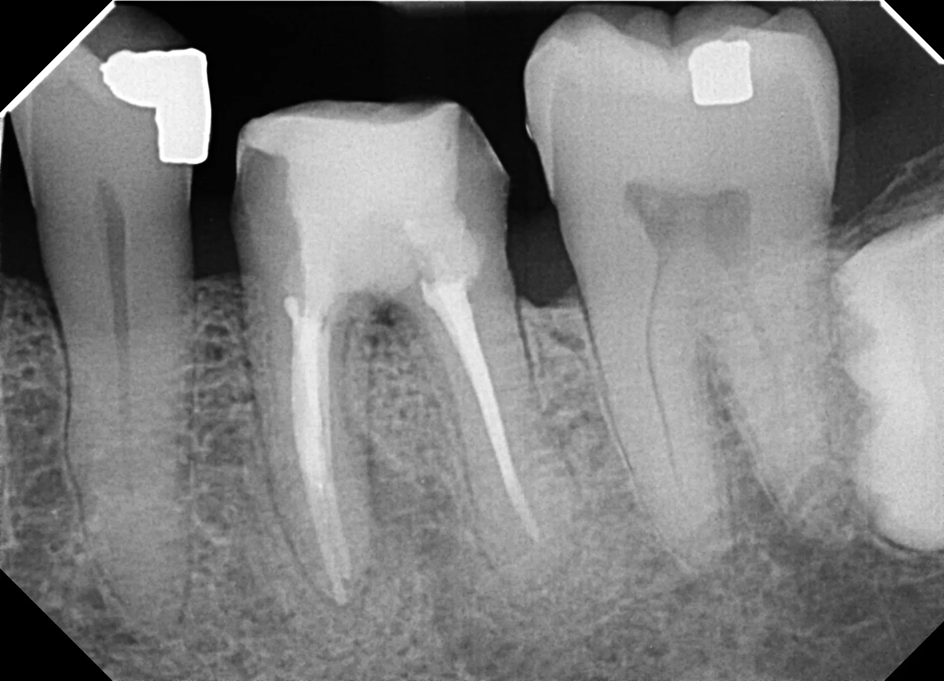
BEFORE AFTER RADIOGRAPH PAIR — CASE 004 — MAXILLARY MOLAR CASE 004 — MAXILLARY MOLAR 9-YEAR FOLLOW-UP The nine-year molar Severe cracking that disrupted sleep with every accidental bite — treated once, bonded once, and pain-free every year since. This is what quality-oriented endodontics means. RESTORATIVE ✦ MODERATE
8-year follow-up radiograph showing significant bone regeneration
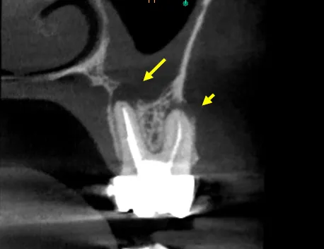
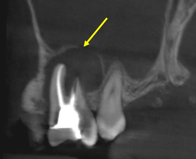
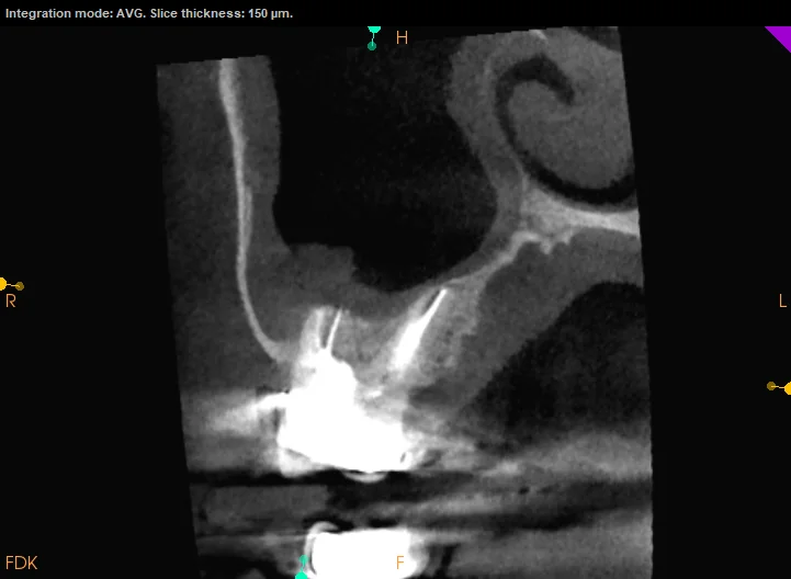
Pre-operative CBCT showing periapical radiolucency into the maxillary sinus
BEFORE AFTER
◂▸
CASE 005 — TOOTH #14 3 MONTHS, SEQUENCED Two diseases, one tooth A 9mm pocket, swelling, and infection reaching toward the sinus — gum disease and nerve death presenting at once. The save depended entirely on treating them in the right order. LESIONS ✦✦ CHALLENGING
CBCT at 12-month recall showing bone regeneration and healing
Pre-operative radiograph showing apical lesion and failed root canal with post
BEFORE AFTER
◂▸
CASE 006 — MANDIBULAR MOLAR UNDER 15 MIN EXTRAORAL Out, repaired, and home again A failing root canal locked behind a large metal post — unreachable from above, impractical from the side. So the tooth came out, was repaired in fifteen minutes, and went back in. INTERESTING ✦✦✦ EXTREME
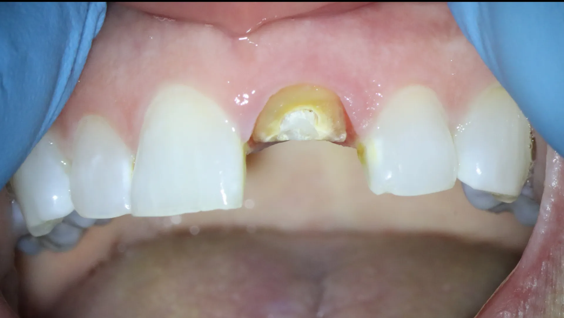
One-year recall — stable tooth with definitive crown restoration
Fractured maxillary central incisor with significant coronal tooth loss
BEFORE AFTER
◂▸
CASE 007 — MAXILLARY CENTRAL INCISOR 90 MINUTES The front tooth that stayed At 62, with a fractured front tooth and a failed root canal beneath it, extraction and an implant seemed inevitable. The patient asked for one more opinion first. RESTORATIVE ✦ MODERATE
BEFORE AFTER RADIOGRAPH PAIR — CASE 008 — TOOTH #14 CASE 008 — TOOTH #14 45-MINUTE CONSULTATION The one that got missed A year of bite pain after a root canal. Monthly adjustments. Two providers, no answers — until a 45-minute consultation and one 3D scan found the canal everyone else had missed. SPECIALIST CONSULTATION ✦ MODERATE
Final radiograph — completed obturation with MTA repair in place
Pre-op CBCT showing furcation perforation with radiolucency
BEFORE AFTER
◂▸
CASE 009 — MANDIBULAR MOLAR 2 APPOINTMENTS Sealing the wrong turn A drill searching for a canal went 2mm past where it should have — straight into the furcation. Three hours later the hole was sealed, and three years later the tooth is still here. MISHAPS ✦✦ CHALLENGING
Periapical radiograph appearing nearly normal despite underlying pathology
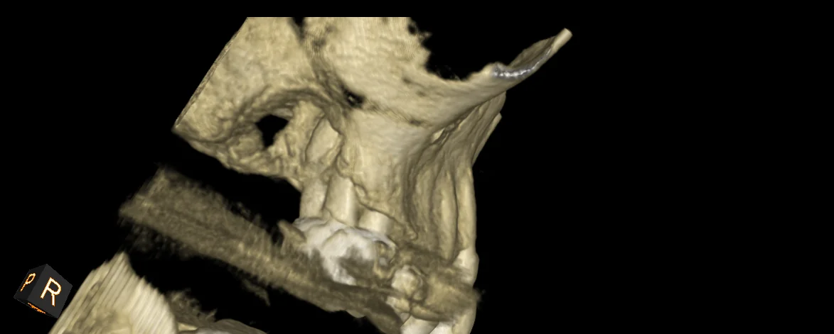
CBCT showing irregular bone borders and cortical plate involvement
BEFORE AFTER
◂▸
CASE 010 — MAXILLARY MOLAR PHASED, MULTIPLE VISITS The ache behind the eye Throbbing tooth pain that peaked every morning, swelling around the eye, antibiotics that did nothing — and a 3D scan whose ragged edges pointed far beyond the tooth. INTERESTING ✦✦ CHALLENGING
BEFORE AFTER RADIOGRAPH PAIR — CASE 011 — LOWER RIGHT QUADRANT CASE 011 — LOWER RIGHT QUADRANT 30-MINUTE EXAM The root canal we didn't do Referred for a root canal after two weeks of ear ache and gum irritation — but every test said the teeth were healthy. The real culprit: a jaw muscle. Total treatment: massage. SPECIALIST CONSULTATION ✦ MODERATE
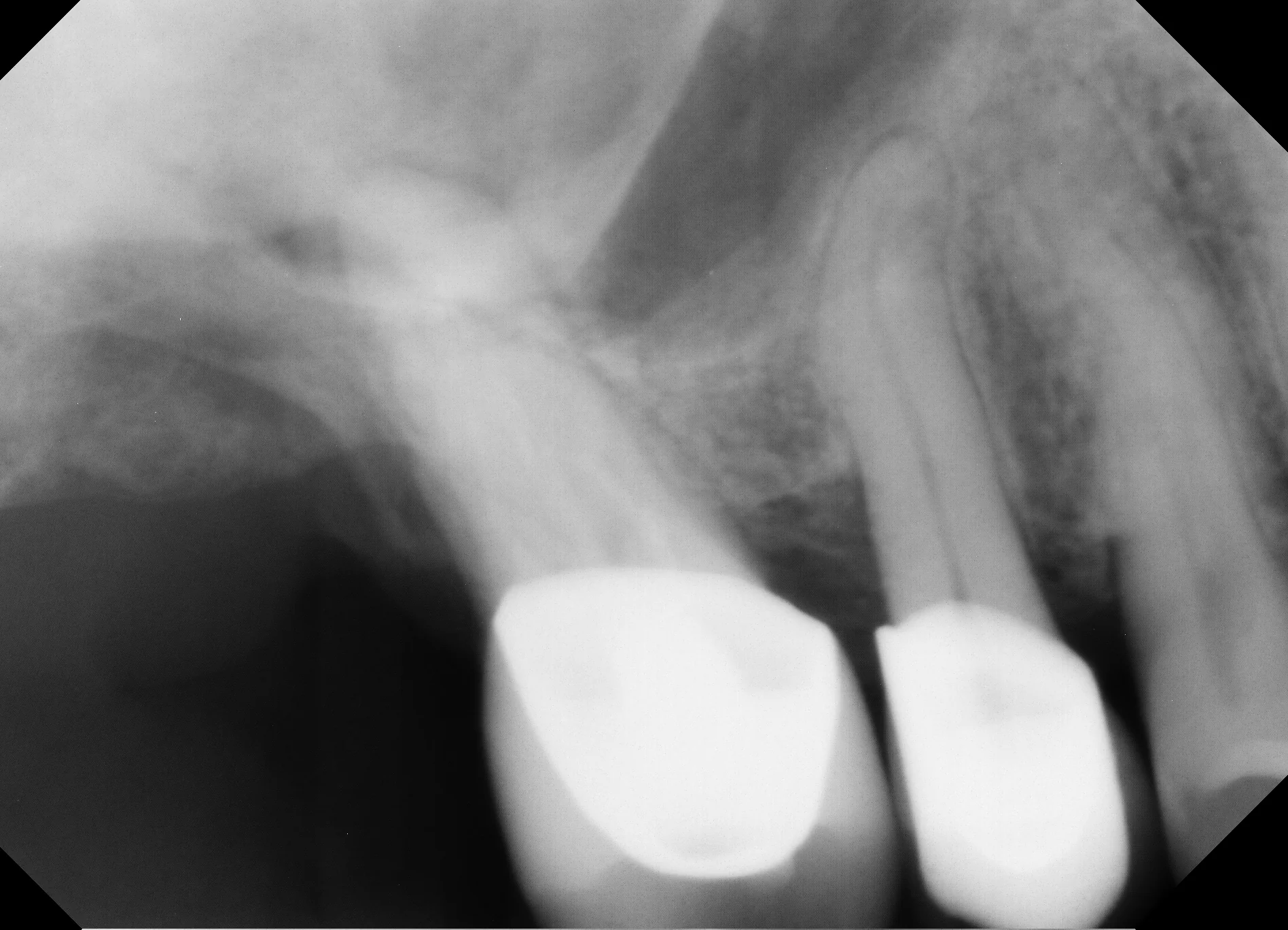
Periapical radiograph of upper right premolar area from the consultation
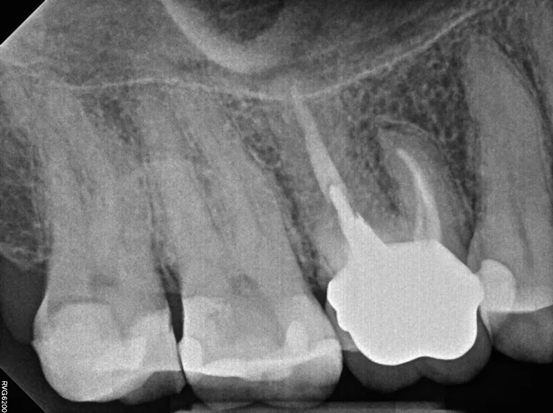
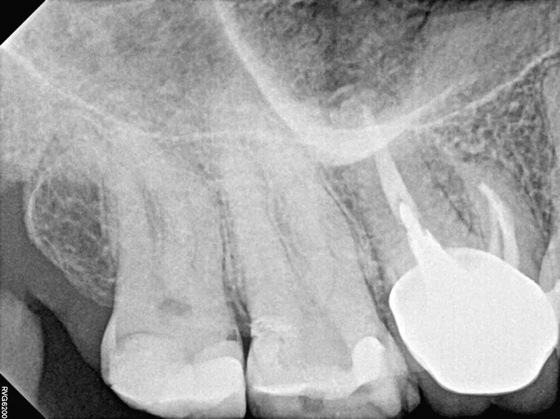
Periapical radiograph of upper right molar region showing subtle changes
BEFORE AFTER
◂▸
CASE 012 — UPPER RIGHT QUADRANT 3-MONTH MONITORING PLAN The hour that changes the answer Chewing pain in the upper right, arriving with holiday-season sinus congestion. Tooth, sinus, or both? Thirty minutes and one 3D scan sorted what months of guessing could not. SPECIALIST CONSULTATION ✦ MODERATE
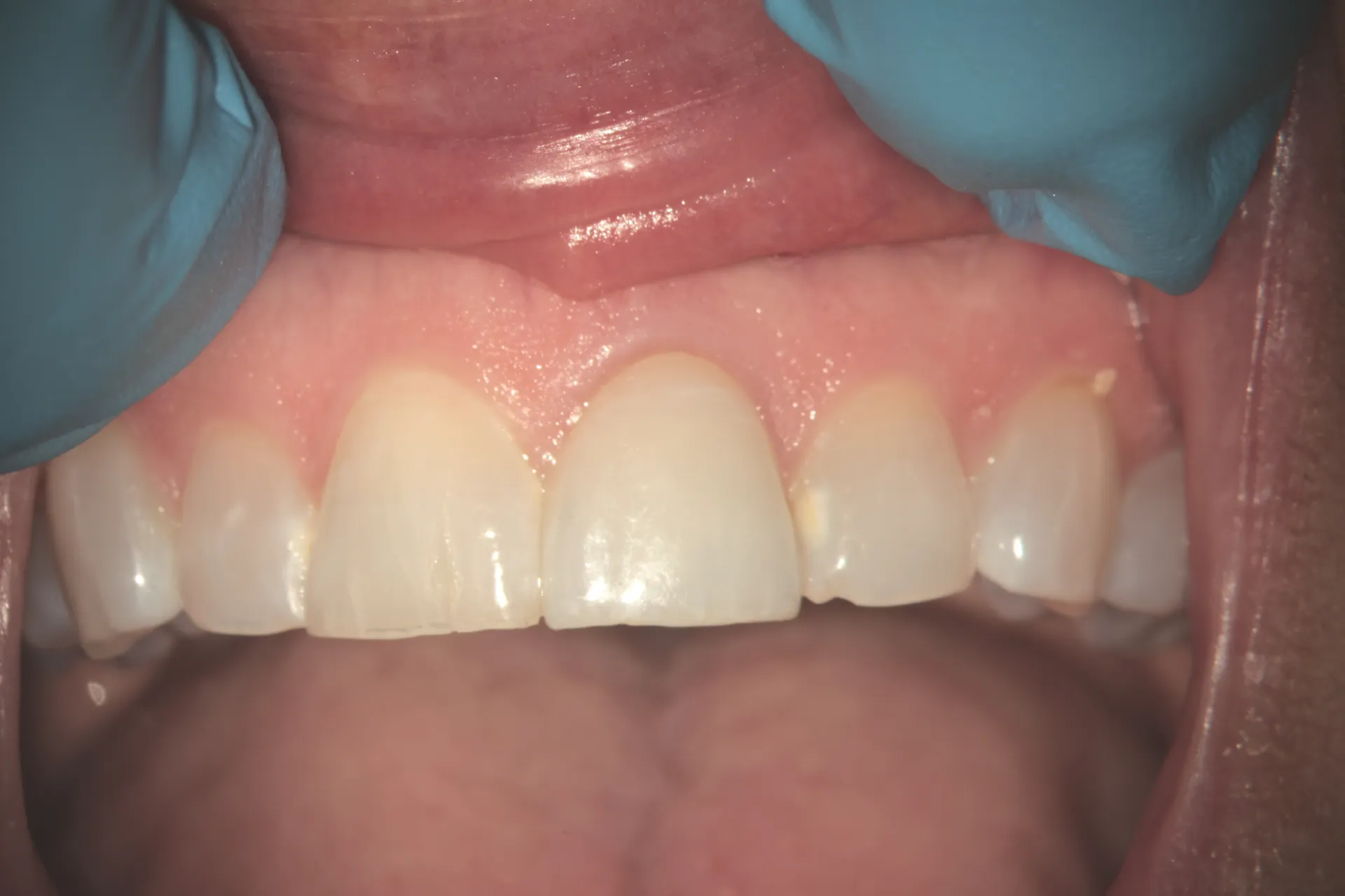
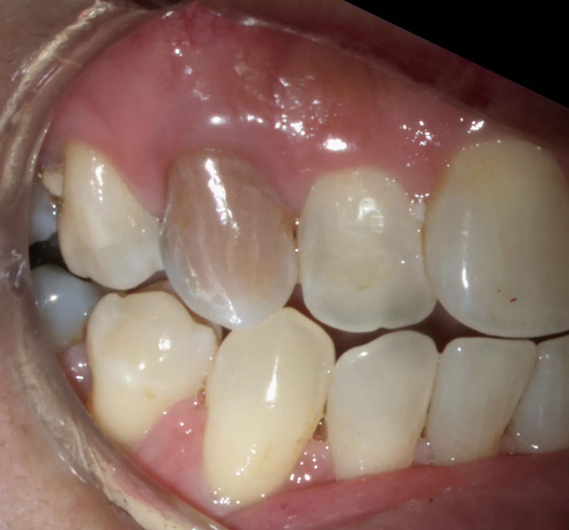
Buccal view after internal bleaching — tooth blending naturally with adjacent teeth
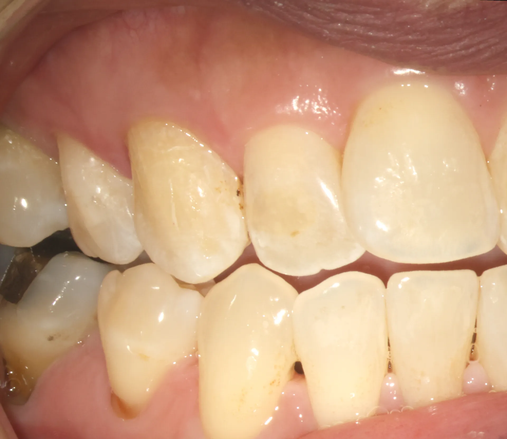
Gray discoloration of maxillary incisor before internal bleaching
BEFORE AFTER
◂▸
CASE 013 — MAXILLARY INCISOR 1 WEEK The original color, restored A front tooth gray for years, whitened from the inside in one week — no crown, no veneer, no tooth reduction. RESTORATIVE ✦ MODERATE
3.4-year recall radiograph — complete resolution of the periapical lesion, tooth in function
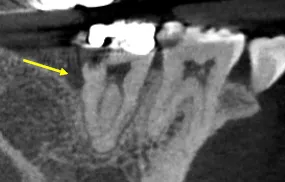
Pre-operative periapical of tooth #19 — prior root canal with persistent periapical pathology
BEFORE AFTER
◂▸
CASE 014 — TOOTH #19 2 VISITS Six problems, one tooth A failing root canal hiding a separated file, a calcified canal, a missed canal, internal resorption, and a crack — quiet and healed 3.4 years after retreatment. MISHAPS ✦✦✦ EXTREME
5-year recall radiograph — bone fill, no recurrent infection, tooth in function
Pre-operative periapical of tooth #3 — prior root canal with persistent pathology and perforation defect
BEFORE AFTER
◂▸
CASE 015 — TOOTH #3 2 PHASES Scheduled for extraction, still here Another provider said the tooth had to go — a perforation, a draining sinus tract, a root canal that had failed. Five years after combined retreatment and surgical repair, it's still doing its job. SURGERY ✦✦✦ EXTREME
SHOWING 15 CASES — THE COLLECTION GROWS
PROVENANCE

Why we show our work.

PROVENANCE Real, not stock Every radiograph is from an actual patient treated in this office.
THE HARD PARTS We show the struggle What made each case difficult, and how we approached it — teaching is proof.
TIME Years, not procedures Recalls at four, six, even fourteen years. Success is measured long after the visit.
HONESTY Including the no's Cases where we advised against treatment — or against ourselves — are here too.
Daily experience
YOUR ONCE-A-YEAR CHALLENGE IS OUR EVERYDAY WORK
Undivided time
OUR SCHEDULE IS BUILT AROUND DIFFICULT ENDODONTICS
Honest reports
YOUR PATIENT COMES BACK TO YOU — WITH THE FULL STORY

Have a case like these?

When standard endodontics isn't enough, we can often save teeth that seem hopeless — and we'll tell you honestly when we can't.

Call (310) 378-8342 Refer a patient