The canal that wasn't there.
Two years of unexplained pain after a "finished" root canal — until 3D imaging revealed a fourth canal, sealed shut by calcification and invisible on every X-ray.
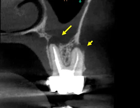
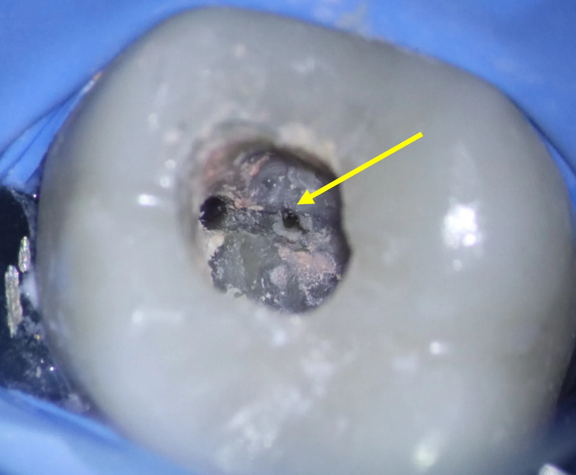
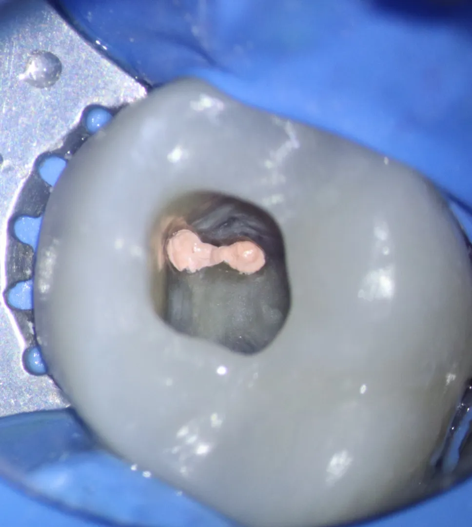
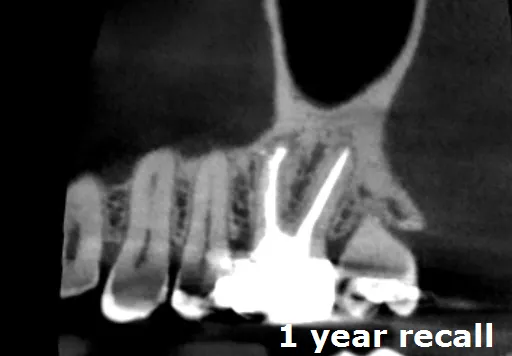
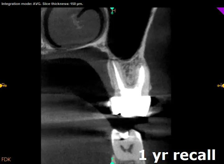
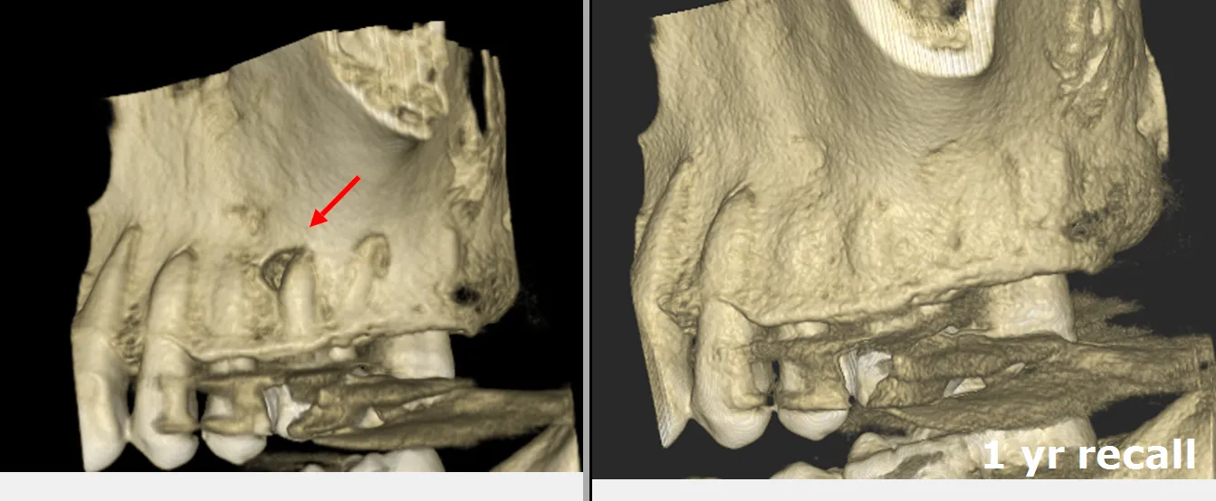
One tooth's journey, in four beats.
A finished tooth, still aching.
A 45-year-old referred by his Manhattan Beach dentist: root canal completed two years earlier, yet the intermittent pain and percussion sensitivity never left. The exam showed nothing wrong.
The map said otherwise.
CBCT imaging told a different story — a completely calcified MB2 canal, invisible on standard X-rays, with infection already forming at the root tip.
Troughing through stone.
Under the microscope, ultrasonic tips carved a pathway through the sclerotic dentin; micro-openers negotiated the canal, and GentleWave irrigation cleaned where needles cannot reach. Two focused hours.
Bone, growing back.
All four canals sealed. At recall the CBCT shows the bone regenerated — the patient asymptomatic two years on, the tooth fully functional.
- ✦ CBCT analysis to pinpoint the MB2 orifice and depth of calcification
- ✦ Surgical microscope at 16× for visualization
- ✦ Ultrasonic troughing (CPR tips) through the sclerotic dentin shelf
- ✦ Size 06–08 micro-openers negotiated to full working length
- ✦ GentleWave multisonic irrigation of the complex anatomy
- ✦ Warm vertical compaction — three-dimensional seal of all four canals
What this case teaches.
More from the collection.
- The C-shaped labyrinth — Severe decay in a lower molar hid a rarity: no separate canals at all, but one continuous C-shaped ribbon of pulp — anatomy that defeats every standard technique.
Pain that outlasted the treatment?
A missed canal is a solvable problem. Start with imaging that sees all of it.