The ache behind the eye.
Throbbing tooth pain that peaked every morning, swelling around the eye, antibiotics that did nothing — and a 3D scan whose ragged edges pointed far beyond the tooth.
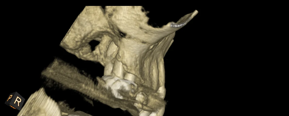
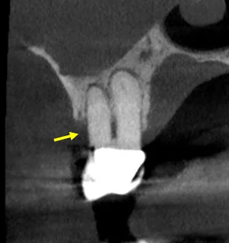
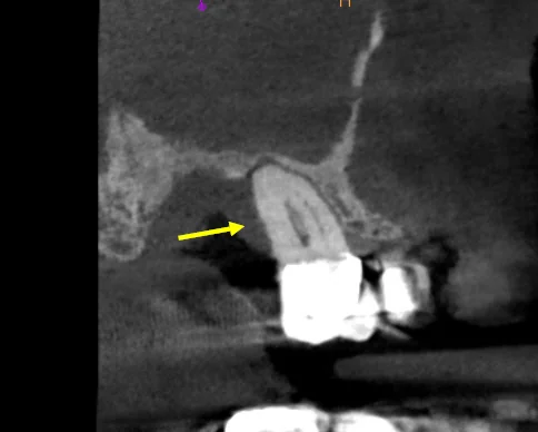
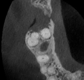
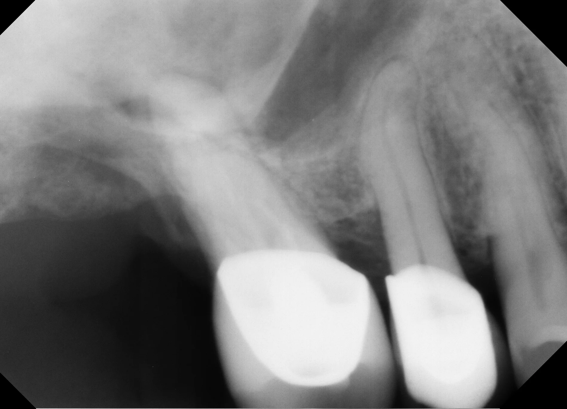
One tooth's journey, in four beats.
Pain that didn't behave.
A 45-year-old Manhattan Beach patient: morning-peaking throbbing in an upper molar, swelling around the eye, and a completed course of antibiotics that changed nothing.
Edges that shouldn't be ragged.
The CBCT showed canal calcification and furcation involvement — but also irregular, ill-defined bone borders eroding the cortical plate. Infections and cysts don't usually look like that.
A test instead of a guess.
Rather than extract on suspicion, the tooth was opened and medicated with calcium hydroxide — a deliberate diagnostic test. Healing would mean infection; non-healing would justify biopsy.
The answer in time.
The tooth didn't heal. Extraction with biopsy confirmed a rare maxillary sinus carcinoma — under 0.2% of sinus pathology — and the patient entered oncology care with a confirmed diagnosis, months earlier than an incidental discovery.
- ✦ History: morning-dominant throbbing pain, eye swelling, antibiotics without effect
- ✦ Exam: 6+ mm distal probing pocket; findings not matching routine infection
- ✦ CBCT: canal calcification, furcation involvement, ragged cortical plate erosion
- ✦ Differential: endo-perio lesion / vertical root fracture / possible sinus malignancy
- ✦ Phased test: tooth opened, calcium hydroxide placed, healing monitored
- ✦ Non-healing → extraction with biopsy → histological diagnosis → prompt oncology referral
What this case teaches.
More from the collection.
- The one that got missed — A year of bite pain after a root canal. Monthly adjustments. Two providers, no answers — until a 45-minute consultation and one 3D scan found the canal everyone else had missed.
- The root canal we didn't do — Referred for a root canal after two weeks of ear ache and gum irritation — but every test said the teeth were healthy. The real culprit: a jaw muscle. Total treatment: massage.
- The hour that changes the answer — Chewing pain in the upper right, arriving with holiday-season sinus congestion. Tooth, sinus, or both? Thirty minutes and one 3D scan sorted what months of guessing could not.
Symptoms that reach beyond the tooth?
Eye swelling, sinus pressure, pain that won't follow the rules — that's a whole-picture workup, not a quick extraction.