The tear beneath the surface.
A well-done root canal that wouldn't heal, a sinus tract that wouldn't close — because the real problem was a torn strip of cementum on the outside of the root, invisible to every X-ray.
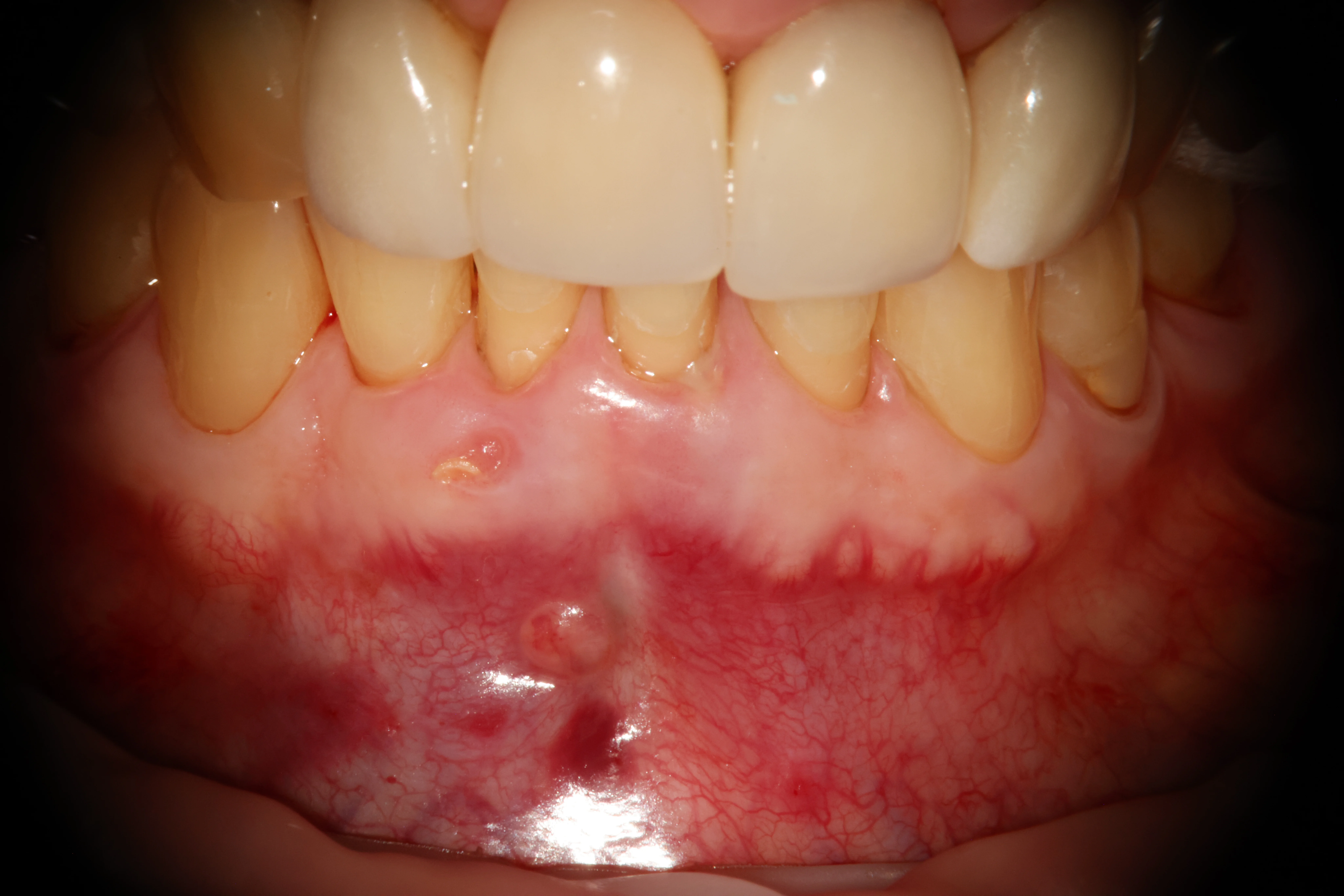
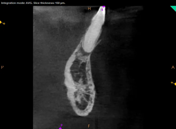
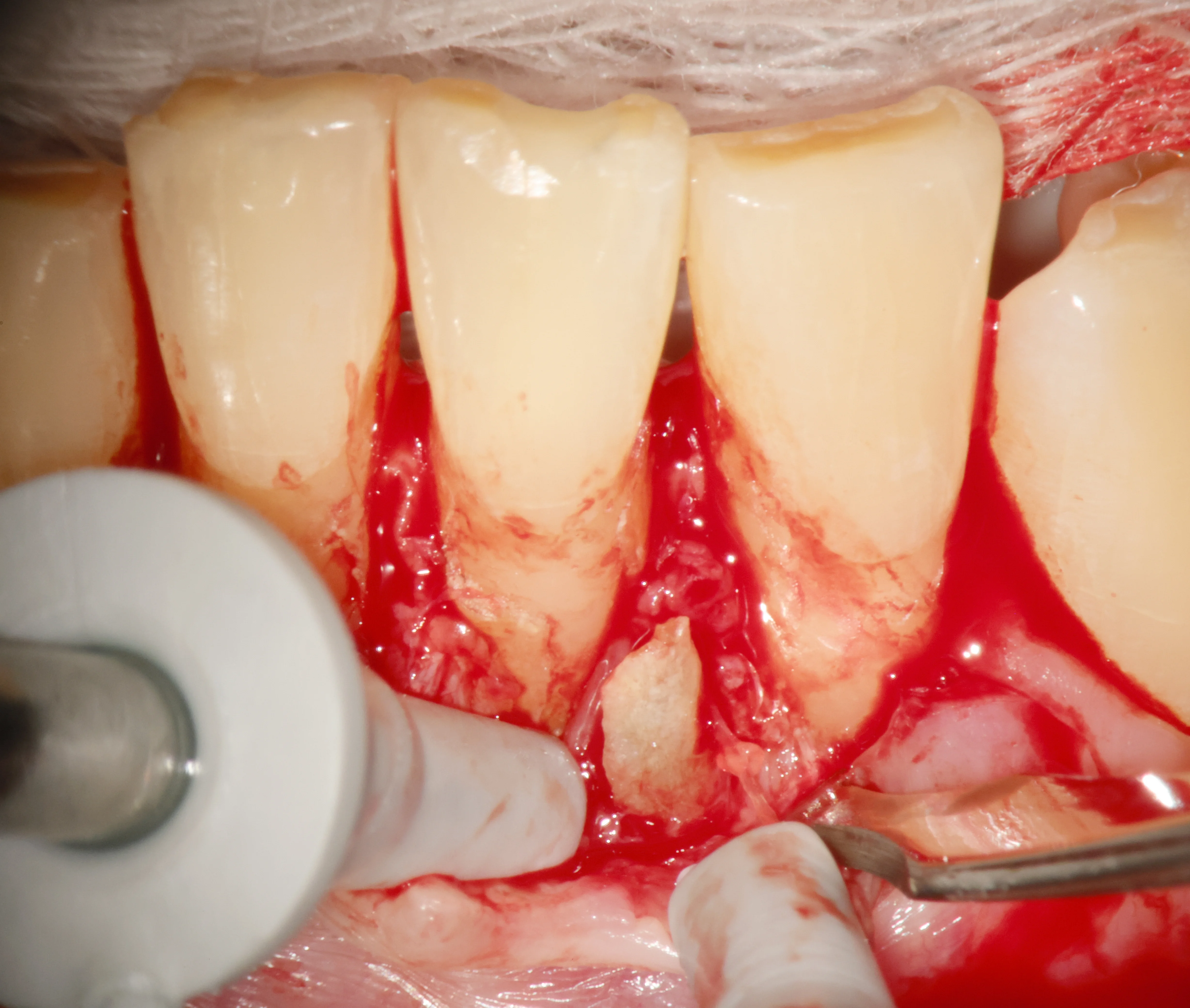
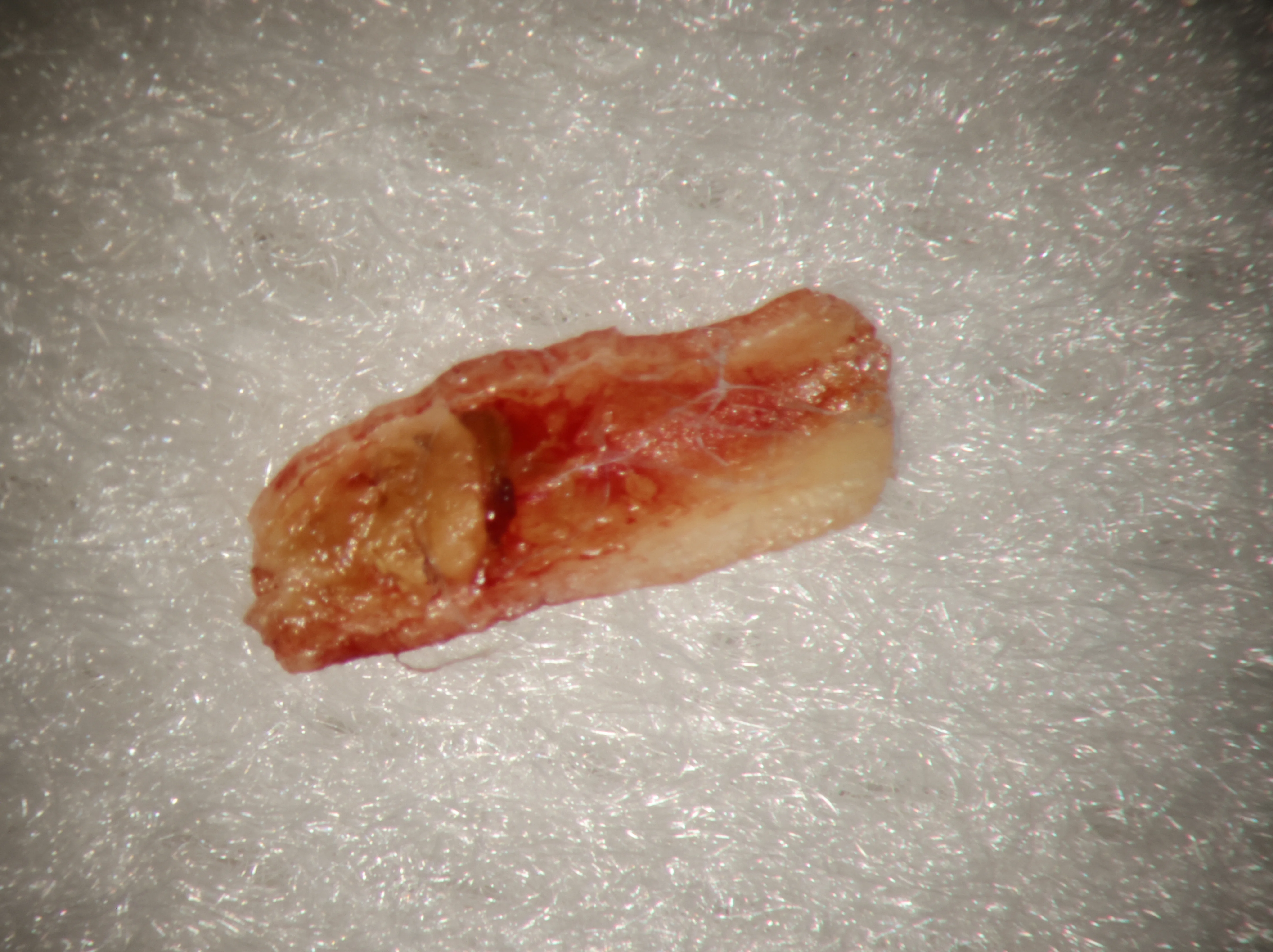
One tooth's journey, in four beats.
Healed on paper, hurting in person.
A Palos Verdes Estates patient months past root canal treatment on two lower front teeth — canals well-filled, yet bite discomfort lingered and infection kept returning through a draining sinus tract.
The scan that didn't add up.
CBCT showed bone loss and a thinning buccal plate — findings the well-obturated canals couldn't explain. Probing ruled out a root fracture. Something external was at work.
Looking directly.
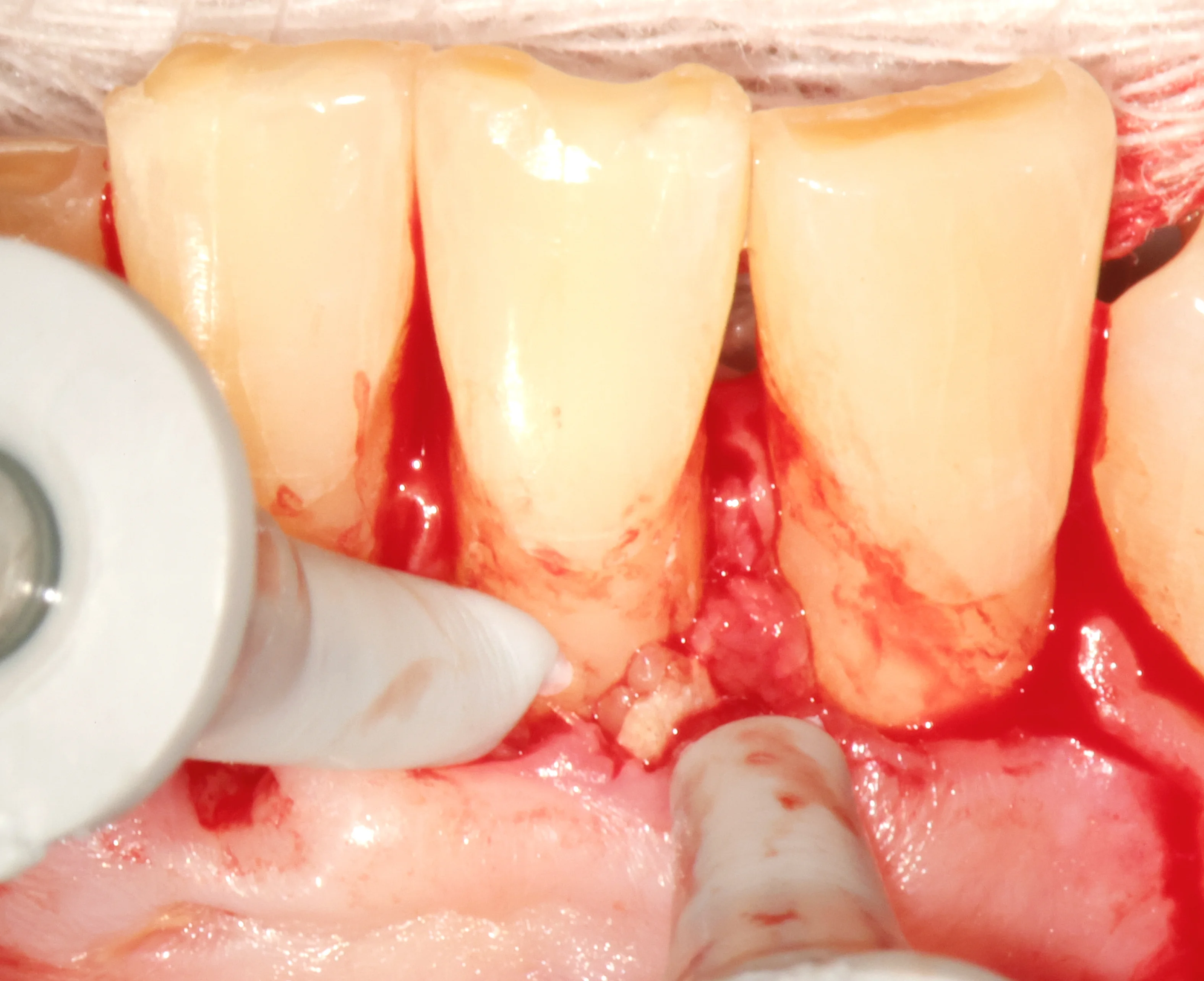
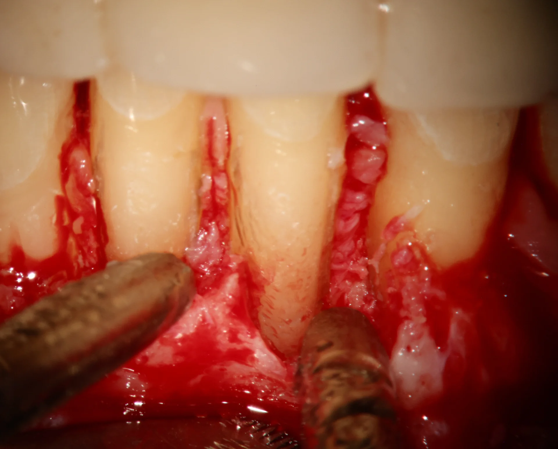
A surgical flap exposed the roots. At 16×, the answer appeared: a 4–5mm vertical cemental tear on #24, a loose fragment harboring a hidden pocket of chronic infection. It was removed and the defect cleaned.
The cause, not the symptom.
The flap was sutured; recovery began immediately. No amount of canal retreatment would ever have reached this — the answer lived on the outside of the root.
- ✦ Full-thickness mucoperiosteal flap under local anesthesia
- ✦ Systematic root-surface inspection at 16× magnification
- ✦ Vertical cemental tear identified — 4–5mm along the root of #24
- ✦ Loose cementum fragment elevated and removed with microsurgical curettes
- ✦ Granulation tissue debrided from the tear defect
- ✦ Flap repositioned and sutured; healing monitored at recall
What this case teaches.
More from the collection.
- Two diseases, one tooth — A 9mm pocket, swelling, and infection reaching toward the sinus — gum disease and nerve death presenting at once. The save depended entirely on treating them in the right order.
A root canal that won't heal?
Retreatment isn't always the answer. Sometimes the cause is where no file can go.