The hour that changes the answer.
Chewing pain in the upper right, arriving with holiday-season sinus congestion. Tooth, sinus, or both? Thirty minutes and one 3D scan sorted what months of guessing could not.
One tooth's journey, in four beats.
Two suspects, one pain.
A Torrance patient with two weeks of chewing discomfort in the upper right — arriving together with nasal congestion and sinus pressure. Dental problem, sinus problem, or both?
Both examined at once.
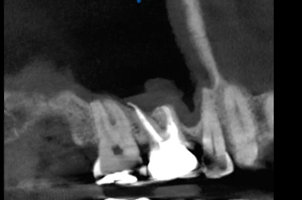
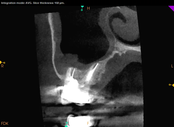
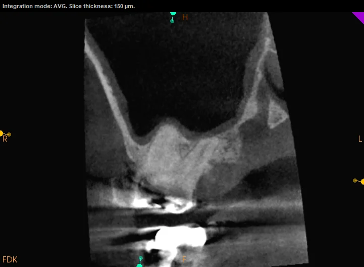
Upper back-tooth roots sit against the sinus floor, so the two mimic each other constantly. CBCT evaluated the teeth and sinus in a single 3D scan — something no standard X-ray can do.
A subtle answer.
The scan found an early endodontic concern in one tooth, invisible on 2D film, plus mild sinus thickening — two overlapping conditions, each needing different management.
Nothing drilled, everything clear.
The plan: monitor the tooth at three months, treat the sinus with the physician, intervene only if the dental finding progresses. An unnecessary root canal avoided — by design.
- ✦ Detailed history: symptom timeline, chewing trigger, relationship to sinus congestion
- ✦ Vitality, percussion, and palpation testing of every tooth in the quadrant
- ✦ CBCT evaluating the teeth and the sinus together, in three dimensions
- ✦ Finding: a subtle endodontic concern in one tooth + mild sinus thickening
- ✦ Plan: monitor at 3 months, coordinate sinus care with the physician
- ✦ Intervene only if the dental finding progresses — not before
What this case teaches.
More from the collection.
- The one that got missed — A year of bite pain after a root canal. Monthly adjustments. Two providers, no answers — until a 45-minute consultation and one 3D scan found the canal everyone else had missed.
- The ache behind the eye — Throbbing tooth pain that peaked every morning, swelling around the eye, antibiotics that did nothing — and a 3D scan whose ragged edges pointed far beyond the tooth.
- The root canal we didn't do — Referred for a root canal after two weeks of ear ache and gum irritation — but every test said the teeth were healthy. The real culprit: a jaw muscle. Total treatment: massage.
Tooth pain with a stuffy nose?
Don't treat the wrong suspect. One consultation looks at the whole picture first.