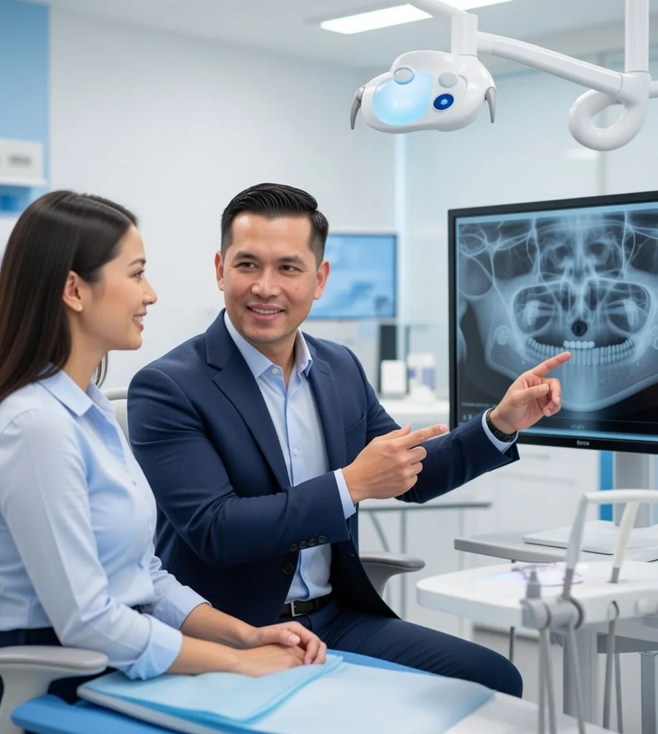
See What Traditional X-Rays Miss
Accurate diagnosis is the foundation of successful treatment. Our CBCT (Cone Beam Computed Tomography) scanner provides three-dimensional visualization, revealing critical information that traditional X-rays cannot show.
Volumetric Imaging
Seconds Scan Time
Complete View
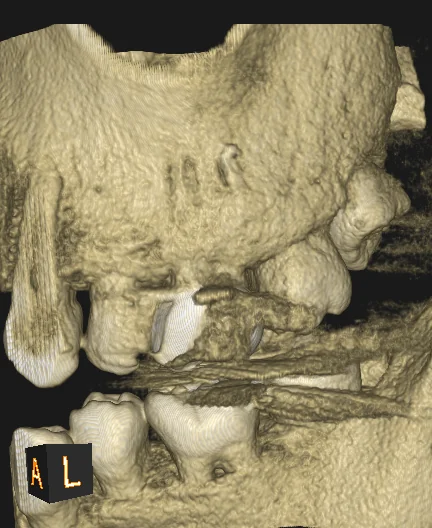
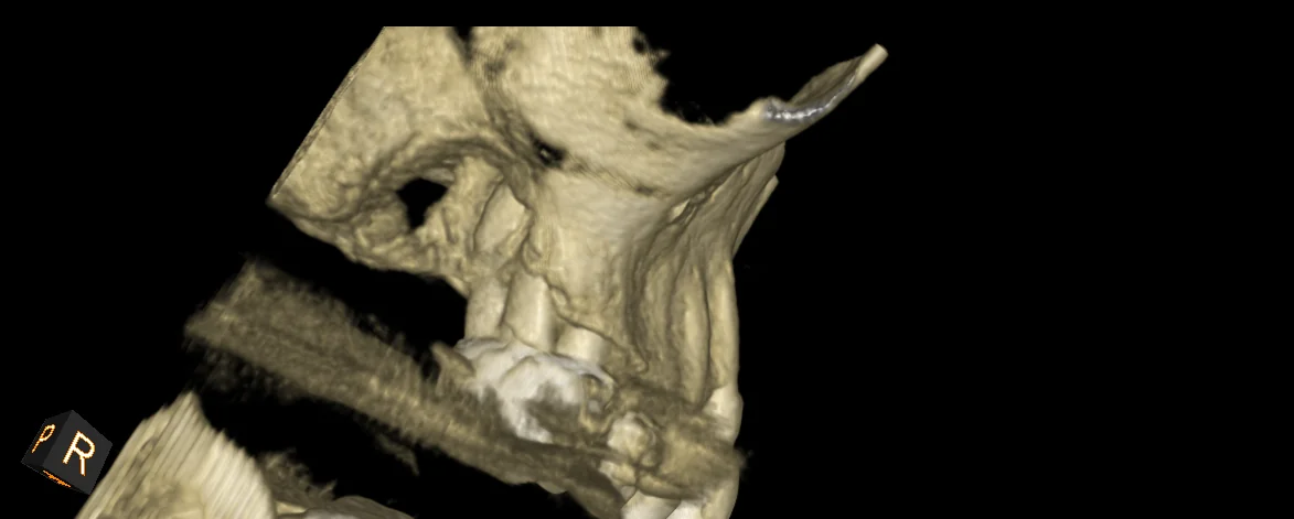
What CBCT Reveals
Hidden Canal Anatomy
- • Extra canals (3-4 when X-rays show 2)
- • C-shaped, lateral, accessory canals
- • Merged/bifurcated roots
- • Calcified canals
Cracks & Fractures
- • Vertical root fractures
- • Craze lines, incomplete fractures
- • Crack extent and direction
- • Root fractures from trauma
Infection & Bone Loss
- • Exact size/location of abscesses
- • Bone destruction around roots
- • Sinus involvement
- • Healing progress monitoring
Previous Dental Work
- • Separated instruments in canals
- • Perforations in tooth structure
- • Over/underfilled root canals
- • Post placement and length
Critical Structures
- • Nerve location to avoid injury
- • Sinus proximity (upper teeth)
- • Adjacent tooth roots
- • Bone thickness for surgery
Treatment Guidance
- • Surgical planning precision
- • Calcified canal navigation
- • Instrument removal strategy
- • Complexity assessment
CBCT vs. Traditional X-rays
| Feature | Traditional X-rays | CBCT 3D Imaging |
|---|---|---|
| Dimension | 2D (flat image) | 3D (volumetric) |
| Hidden Canals | Often missed | Clearly visible |
| Cracks/Fractures | Difficult to detect | Clearly shown |
| Infection Size | Estimated | Precisely measured |
| Overlapping Structures | Can obscure findings | No overlap |
| Diagnostic Accuracy | Good for basic cases | Superior for complex cases |
The Scanning Process
Quick scan duration
Immediate results
Open scanner design
Stand or sit in scanner
Rest chin on support
Remain still 20-40 seconds
Images appear instantly
Safety & Radiation
Low Radiation Dose
- Significantly less radiation than medical CT scans
- Similar to full mouth X-ray series or slightly more
- Focused beam exposes only the area being imaged
- Latest technology minimizes necessary dosage
We follow ALARA principle (As Low As Reasonably Achievable) - only used when 3D information will change diagnosis or treatment.
When We Use CBCT
Recommended For:
- ✓ Unclear diagnosis from traditional X-rays
- ✓ Complex anatomy suspected
- ✓ Retreatment needed
- ✓ Surgical planning required
- ✓ 3D visualization provides clear benefit
Insurance & Cost
CBCT imaging is typically billed separately from treatment and covered by most dental insurance when medically necessary.
Typical cost range
Often covered by insurance
We check coverage first
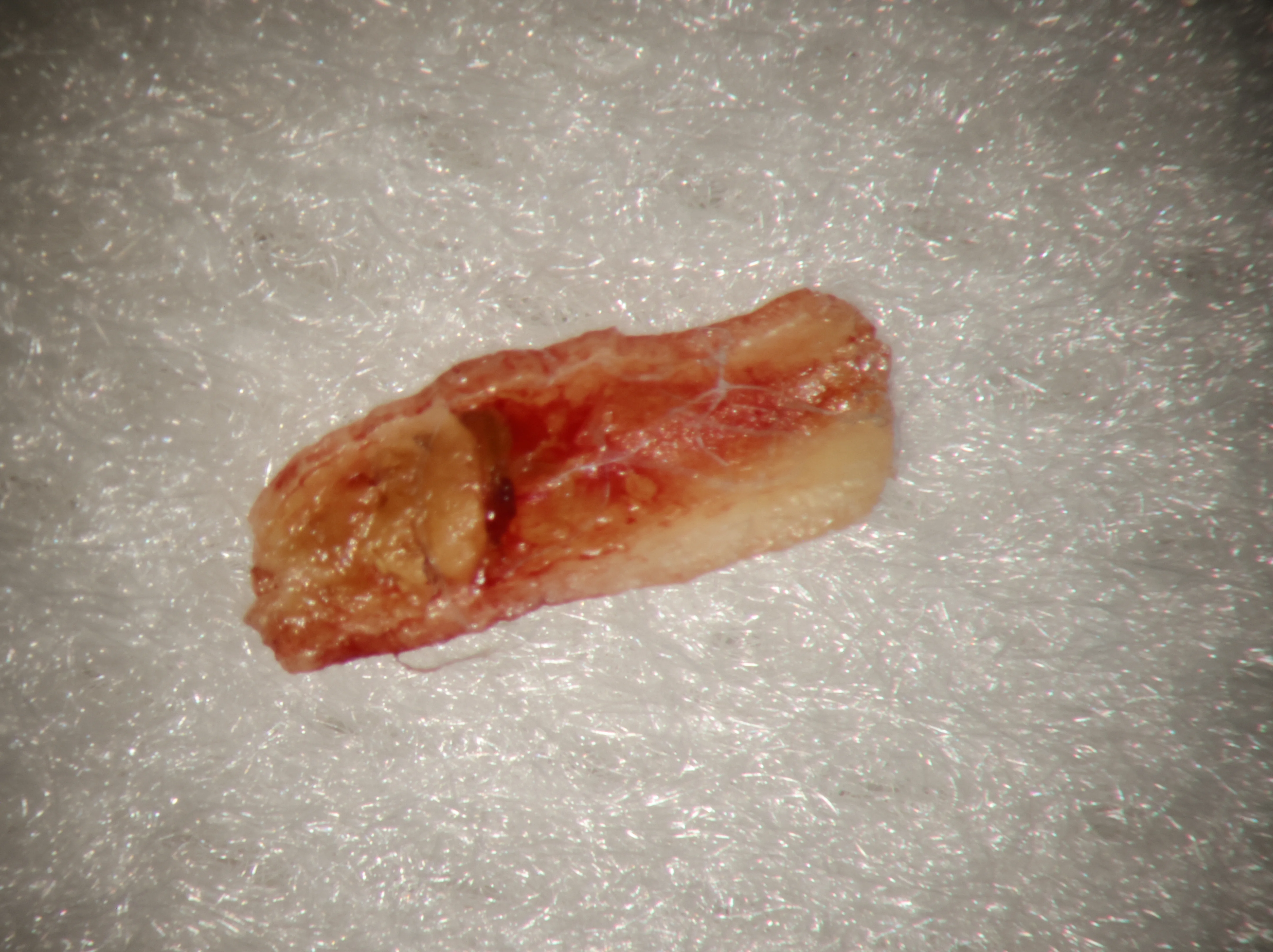
Oral Pathology Diagnosis
One of the most valuable — and often overlooked — uses of CBCT imaging is identifying oral pathology that doesn’t show up on regular X-rays.
When a patient comes in with unexplained pain, swelling, or a lesion that doesn’t quite make sense, our 3D imaging can reveal what’s really going on. We’re talking about things like:
Periapical Cysts and Granulomas — These fluid-filled sacs or inflammatory tissue masses form around infected root tips. On a flat X-ray, they look similar. On CBCT, we can see the exact size, shape, and relationship to surrounding structures — which changes how we approach treatment.
Odontogenic Keratocysts — These aggressive cysts can grow quietly inside the jawbone. CBCT shows their true extent, helping oral surgeons plan the right intervention. We’ve caught a few of these over the years that would have been missed on standard imaging.
Resorption — Both internal resorption (the tooth dissolving from the inside) and external resorption (the root being eaten away from the outside) are much easier to diagnose and measure in 3D. Knowing the exact extent of resorption determines whether a tooth can be saved.
Dentigerous Cysts — Associated with impacted or unerupted teeth, these show up clearly on CBCT with their full relationship to nerves, sinuses, and adjacent teeth.
When we spot something on a scan that falls outside the scope of endodontics, we refer promptly to the appropriate specialist — whether that’s an oral surgeon, periodontist, or oral pathologist. Our job is to catch it. And 3D imaging makes us much better at catching it.
Advanced Imaging for Torrance and the South Bay
CBCT 3D imaging is one example of how our Torrance endodontic office uses the latest diagnostic technology. Combined with surgical microscopy and Dr. Phan’s expertise, advanced imaging means more accurate diagnoses and better treatment outcomes for patients across the South Bay.
See your teeth like never before. Schedule your consultation.